
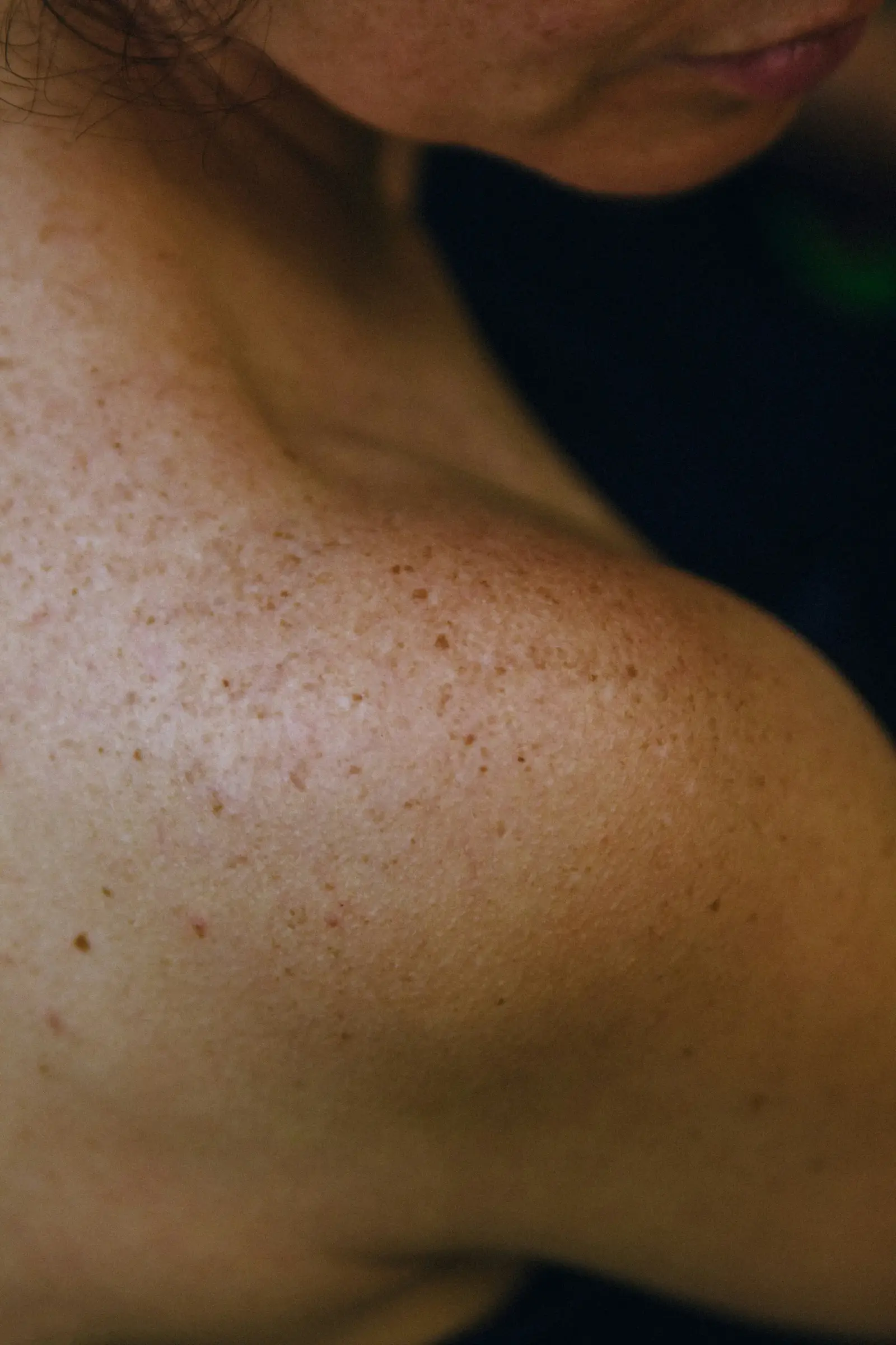
Pre-Cancerous Lesions
Pre-cancerous skin changes can be identified and treated before they progress. A professional exam helps distinguish actinic keratoses from other spots.
About the Condition
Your provider examines areas of concern and may use a dermatoscope or biopsy when a spot needs a closer look. The plan depends on the lesion, its location, and your medical history.
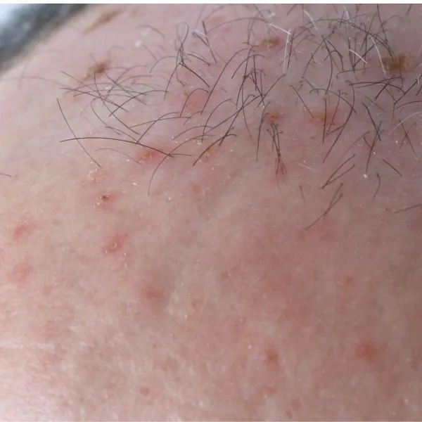
Causes
Actinic keratoses are commonly associated with cumulative ultraviolet exposure from the sun or tanning beds. Sun protection and routine skin exams can help support early detection.
Symptoms To Watch For
- A rough, scaly patch that may feel easier to notice than to see
- A dry or crusted spot on sun-exposed skin
- A lesion that persists or returns after healing
- A firm or raised area that changes over time
Who’s At Risk
Risk can increase with years of sun exposure, repeated sunburns, fair skin, and a history of actinic keratoses or skin cancer.
Evaluation & Treatment Options
Skin Examination
A provider assesses the spot and checks surrounding skin for other changes.
Cryotherapy
A quick in-office treatment may be used for selected individual lesions.
Topical Treatment
Prescription therapy may be recommended for selected areas or multiple lesions.
Photodynamic Therapy
Light-activated treatment can be an option for selected precancerous lesions.
Outlook & Living With It
Early evaluation helps your provider choose the right treatment and monitor for changes. Regular skin exams and sun protection are important parts of ongoing care.
Frequently Asked Questions
What is an actinic keratosis?
It is a rough, scaly area caused by sun damage that a provider can evaluate and treat when appropriate.
Can a pre-cancerous spot become skin cancer?
Some precancerous lesions can progress, so persistent or changing spots should be evaluated by a dermatology provider.
How can I reduce future sun damage?
Use broad-spectrum sunscreen, protective clothing, and shade, and avoid tanning beds. Your provider can recommend a skin-check schedule for your history.